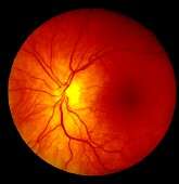
MS中的光学相干断层扫描IDS脑萎缩

神经节细胞+内丛状层(GCIP)萎缩的比率反映了多发性硬化症(MS)的全脑萎缩,根据最佳相干断层扫描(OCT)的测量,7月18日在线发表在《健康日》杂志上的一项研究神经病学年鉴。
In order to validate the utility of OCT as an indicator of neuronal tissue damage in patients with MS, Shiv Saidha, M.B.B.Ch., from Johns Hopkins University in Baltimore, and colleagues examined whether atrophy of specific retinal layers and brain substructures are associated over time. They performed biannual cirrus high definition OCT in 107 patients with MS.
研究人员观察到相关在GCIP和全脑,灰质(GM),白质(WM)和丘脑萎缩的速率之间。GCIP和全部有更强烈的相关性 -脑萎缩进展性MS与复发-缓解性MS (RRMS)的比率(r = 0.67;P < 0.001, r = 0.33;P = 0.007)。在RRMS中,GCIP和全脑(GM和WM)萎缩之间的相关性随着逐步细化而逐渐增加,以排除ON(有视神经炎病史的眼睛)影响;除去眼睛,再除去患者,相关性分别增加到0.45和0.60,与效果矫正相一致。病变积累率与GCIP和内层核层萎缩率相关。
作者写道:“我们的研究结果支持OCT用于临床监测和作为调查试验的结果。”
若干作者向制药和生物技术行业披露了财务关系。
版权©2015每日健康。保留所有权利。


用户评论