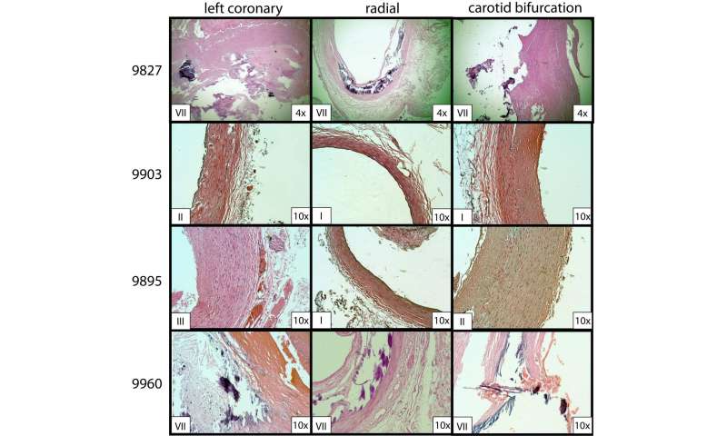
该图显示了根据美国心脏协会(AHA)对动脉粥样硬化分级所使用的组织学分级表。该图的目的是给出标准,并解释为什么各部分被分配特定的等级。切片用苏木精和伊红染色。星形表示血管腔。水平的面板代表研究中观察到的每个等级,I-VII(不包括VI)。垂直的柱子描述了在不同放大倍率下相同的剖面。I级:仅存在孤立的巨噬细胞泡沫细胞(箭头突出显示)。这是颈外动脉的一部分。II级:巨噬细胞泡沫细胞呈层状,椭圆形包围。这是胫骨后动脉的一部分。III级:可见细胞外脂质小池,如矩形所示。 This is a section of the vertebral artery. Grade IV: exhibits the presence of a large confluent extracellular lipid pool, indicated by the rectangle. This is a section of the femoral artery. Grade V: has apparently thickened fibromuscular tissue layer, appreciated by the line with brackets. An extracellular lipid pool is also present here, indicated by the rectangle. This is a section of the internal carotid artery. Grade VII: calcification predominates the tunica of the vessel wall, indicated by the green asterisk. This is a section of the radial artery. Credit: Christopher Hoehmann, NYIT Medical Student
“外周动脉可能是冠状动脉疾病的可靠指标”,作者是Brian L. Beatty博士和Bennett Futterman医学博士,他们都是NYITCOM的解剖学副教授,以及Christopher Hoehmann,三年级学生医科学生在那里。在他们的研究中,作者研究了48具尸体的动脉,以确定动脉粥样硬化的风险因素,从每具捐赠的尸体中取样了13个动脉段,包括颈动脉、中央动脉和外周动脉的段。
 to assign grades of atherosclerosis. The purpose of this figure is to give criteria and explain why sections were assigned specific grades. Sections are stained in hematoxylin and eosin. Stars indicate the vessel lumen. Horizontal panels represent each grade observed in the study, I-VII (excluding VI). The vertical columns depict the same sections at different magnifications. Grade I: only isolated macrophage foam cells are present (highlighted by the arrow). This is a section of the external carotid artery. Grade II: macrophage foam cells are apparent here in layers, encircled by the elliptical shapes. This is a section of posterior tibial artery. Grade III: has small pools of extracellular lipids visible, depicted by the rectangle. This is a section of the vertebral artery. Grade IV: exhibits the presence of a large confluent extracellular lipid pool, indicated by the rectangle. This is a section of the femoral artery. Grade V: has apparently thickened fibromuscular tissue layer, appreciated by the line with brackets. An extracellular lipid pool is also present here, indicated by the rectangle. This is a section of the internal carotid artery. Grade VII: calcification predominates the tunica of the vessel wall, indicated by the green asterisk. This is a section of the radial artery. Credit: Christopher Hoehmann, NYIT Medical Student")