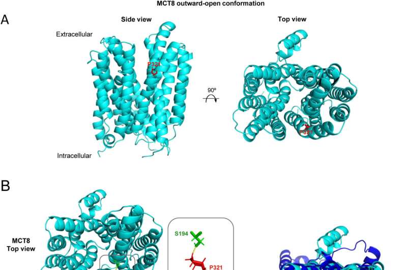
MCT8向外开放构象同源模型的三级结构。(A)基于人MCT1 (PDB: 6LYY)向外开放构象的冷冻电镜结构,使用Robetta服务器预测MCT8结构。对模型进行了改进,以消除不精确预测的残差(>5 Å的误差)。用PyMOL软件生成MCT8结构的侧面(左)和顶部(右)卡通视图。在2例AHDS患者中突变的脯氨酸321残基呈红色棒状。(B) MCT8(浅蓝色)和MCT8- p321l(深蓝色)模型的3D比对显示突变体中TMHs之间的距离缩短。P321L突变改变MCT8的氨基酸相互作用网络。两个模型之间TMHs位置的变化用红色箭头表示(右图)。MCT8-P321L结构预测如a所述,采用Pymol软件进行三维比对。P321L突变改变MCT8的氨基酸相互作用网络(左图)。 While P321 residue only interacts with S194 from TMH1, the P321L mutant also interacts with I197 from TMH1 and W431, L434, and V435 from TMH8. Intra-protein interactions of MCT8 and MCT8-P321L were assessed by RINGS 2.0 and the predicted amino acidic interaction networks were represented by Pymol. Residues are shown as colored sticks. (C) A comparison of the structure of the inner pocket of MCT8 (yellow) and MCT8-P321L (green) shows a dramatic change in the shape and size of the pocket. MCT8 (light blue) and MCT8-P321L (dark blue) are shown as cartoons with the inner cavity represented as a surface in yellow and green respectively. Hormone influx sense was represented by blue arrows. Triiodothyronine (T3) structure was represented as yellow sticks. CASTp server and PyMOL were used for substrate-binding pocket volume analysis and image generation. Credit:疾病神经生物学(2022)。DOI: 10.1016 / j.nbd.2022.105896
. DOI: 10.1016/j.nbd.2022.105896")